Experimental Procedures
Cell Preparation:
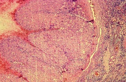
Mixed luteal cells were made from four bovine corpora lutea. Luteal tissue was digested by the use of collagenase type 1 enzyme solution. Cells were washed 3 times with 1x PBS and centrifuged at 300 xg for 5 minutes at 25ºC. Cells were then incubated in T-75 flasks which contained Ham’s F-12 medium with 5% fetal calf serum, insulin, transferrin (5 µg/ml), selenium (5 ng/ml), 100 U/ml penicillin, 0.1 mg/ml streptomycin, and 0.25 mg/ml amphotericin B in an atmosphere of humidified 95% air and 5% CO2 at 37ºC. When confluence was reached, cells either remained untreated, control, or treated with fish oil at 3 x 10⁻² (vol/vol) for a period of 48 hours. 10mM of Beta methyl cyclodextrin (βMCD), which is a reagent known to remove cholesterol, was used as a negative control.
Mixed luteal cells were made from four bovine corpora lutea. Luteal tissue was digested by the use of collagenase type 1 enzyme solution. Cells were washed 3 times with 1x PBS and centrifuged at 300 xg for 5 minutes at 25ºC. Cells were then incubated in T-75 flasks which contained Ham’s F-12 medium with 5% fetal calf serum, insulin, transferrin (5 µg/ml), selenium (5 ng/ml), 100 U/ml penicillin, 0.1 mg/ml streptomycin, and 0.25 mg/ml amphotericin B in an atmosphere of humidified 95% air and 5% CO2 at 37ºC. When confluence was reached, cells either remained untreated, control, or treated with fish oil at 3 x 10⁻² (vol/vol) for a period of 48 hours. 10mM of Beta methyl cyclodextrin (βMCD), which is a reagent known to remove cholesterol, was used as a negative control.
Fractionation:
48 hours later, the supernatant of the T-75 flasks were removed. The cells were then trypsinized and centrifuged to form a cell pellet. The cell pellet was transferred to 0.66mL 0.5M Na2CO3 for cell lysis. These samples were then hand homogenized using a douncer for 10 strokes. Lysis underwent further homogenization 30 times with a 27 gauge syringe. The samples then experienced either 0% or 10% ultrasonic homogenization. Equal parts of 0.66mL 90% sucrose 1M MES (20% glycerol, 150 mM NaCl, 2 mM EDTA, 25 mM MES; pH 6.5) solution was incorporated to create a final volume of 1.32mL 45% sucrose solution at 0.5M MES and 250mM Na2CO3 concentrations. Additional layers of 1.32mL 35% sucrose and 1.32mL 5% sucrose were meticulously added to the 45% sucrose cell lysis solution which formed interfaces at each layer in 12mm x 75mm ultracentrifugation tubes. Treatments were then ultracentrifuged for a period of 24 hours at 250,000 xg at 4ºC utilizing a SW-60 Beckman rotor in-vacuum.
Protein Isolation:
Once centrifugation was finished, 400 µL aliquots were removed beginning from the top of the ultracentrifugation tube and then pipette into 10 marked 1.7mL eppe tubes. Ten equal fractions were collected and an aliquot of each fraction was subjected to SDS-PAGE and western blotting. A 200 µL aliquot was put in 10 new eppe tubes from each sample. Next, 200µL of trichloroacetic acid (TCA) was combined to each sample and then protein was isolated overnight at 4ºC. These samples were centrifuged at 20,000 xg for 10 minutes at 4ºC. They were then washed 2 times in ice cold acetone. Samples were reconstituted in 15µL 1x loading buffer to prepare them for western blot analysis.
Western Blot Analysis:
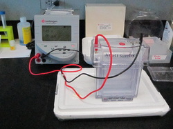
Pre-cast 10% Bis-Tris NuPage western blot gels were loaded with both 5 µL pre-stain biotinylated ladder and horse radish peroxide (HRP) ladder. Then fractions 1-10 of the gel were loaded with 15 µL of isolated protein samples which had already been prepared. The gel was then run for a period of 1 hour and 15 minutes at 140V. After, proteins from the gel were transferred to PvDF membranes through the use of a transfer module at 25V for 1 hour and 15 minutes. These membranes were washed 5 times with 1x TBS-t. They were then blocked with 5% non-fat dry milk for 1 hour. Membranes were probed with primary antibodies for flotillin and caveolin, two proteins which are associated with lipid microdomains. The primary antibodies were allowed to remain overnight at 4ºC. Membranes were washed 5 times in 1x TBS-t. A secondary anti-rabbit antibody was then added for 1 hour at 25ºC. After 1 hour, ultrasensitive chemoluminescence was applied which allowed for the visualization of flotillin and caveolin through the use of a Bio-Rad Versa-Doc imager. Protein density for flotillin and caveolin was quantitated using ImageJ software.
48 hours later, the supernatant of the T-75 flasks were removed. The cells were then trypsinized and centrifuged to form a cell pellet. The cell pellet was transferred to 0.66mL 0.5M Na2CO3 for cell lysis. These samples were then hand homogenized using a douncer for 10 strokes. Lysis underwent further homogenization 30 times with a 27 gauge syringe. The samples then experienced either 0% or 10% ultrasonic homogenization. Equal parts of 0.66mL 90% sucrose 1M MES (20% glycerol, 150 mM NaCl, 2 mM EDTA, 25 mM MES; pH 6.5) solution was incorporated to create a final volume of 1.32mL 45% sucrose solution at 0.5M MES and 250mM Na2CO3 concentrations. Additional layers of 1.32mL 35% sucrose and 1.32mL 5% sucrose were meticulously added to the 45% sucrose cell lysis solution which formed interfaces at each layer in 12mm x 75mm ultracentrifugation tubes. Treatments were then ultracentrifuged for a period of 24 hours at 250,000 xg at 4ºC utilizing a SW-60 Beckman rotor in-vacuum.
Protein Isolation:
Once centrifugation was finished, 400 µL aliquots were removed beginning from the top of the ultracentrifugation tube and then pipette into 10 marked 1.7mL eppe tubes. Ten equal fractions were collected and an aliquot of each fraction was subjected to SDS-PAGE and western blotting. A 200 µL aliquot was put in 10 new eppe tubes from each sample. Next, 200µL of trichloroacetic acid (TCA) was combined to each sample and then protein was isolated overnight at 4ºC. These samples were centrifuged at 20,000 xg for 10 minutes at 4ºC. They were then washed 2 times in ice cold acetone. Samples were reconstituted in 15µL 1x loading buffer to prepare them for western blot analysis.
Western Blot Analysis:
Pre-cast 10% Bis-Tris NuPage western blot gels were loaded with both 5 µL pre-stain biotinylated ladder and horse radish peroxide (HRP) ladder. Then fractions 1-10 of the gel were loaded with 15 µL of isolated protein samples which had already been prepared. The gel was then run for a period of 1 hour and 15 minutes at 140V. After, proteins from the gel were transferred to PvDF membranes through the use of a transfer module at 25V for 1 hour and 15 minutes. These membranes were washed 5 times with 1x TBS-t. They were then blocked with 5% non-fat dry milk for 1 hour. Membranes were probed with primary antibodies for flotillin and caveolin, two proteins which are associated with lipid microdomains. The primary antibodies were allowed to remain overnight at 4ºC. Membranes were washed 5 times in 1x TBS-t. A secondary anti-rabbit antibody was then added for 1 hour at 25ºC. After 1 hour, ultrasensitive chemoluminescence was applied which allowed for the visualization of flotillin and caveolin through the use of a Bio-Rad Versa-Doc imager. Protein density for flotillin and caveolin was quantitated using ImageJ software.
