Results
In this experiment,
there were two types of βMCD, control, and fish oil. These types were such that
one experienced 0% ultrasonic homogenization and the other experienced 10%
ultrasonic homogenization. The reason behind the two different power levels of
ultrasonic homogenization was to achieve the best possible results. This was a
factor taken into account in the separation of results.
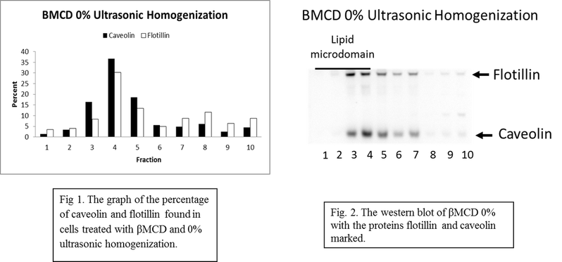
βMCD 0% ultrasonic homogenization had the highest percentage of caveolin and flotillin in fractions 1-4. Fractions 5-10 had lower levels of the two proteins. However, fraction 5 had a significant amount of caveolin and flotillin (Fig. 1). The western blot shows that the greatest amount of proteins was found within the upper fractions such as 2-4. Fraction 5 had a large amount of both proteins (Fig. 2).
βMCD 0% ultrasonic homogenization had the highest percentage of caveolin and flotillin in fractions 1-4. Fractions 5-10 had lower levels of the two proteins. However, fraction 5 had a significant amount of caveolin and flotillin (Fig. 1). The western blot shows that the greatest amount of proteins was found within the upper fractions such as 2-4. Fraction 5 had a large amount of both proteins (Fig. 2).
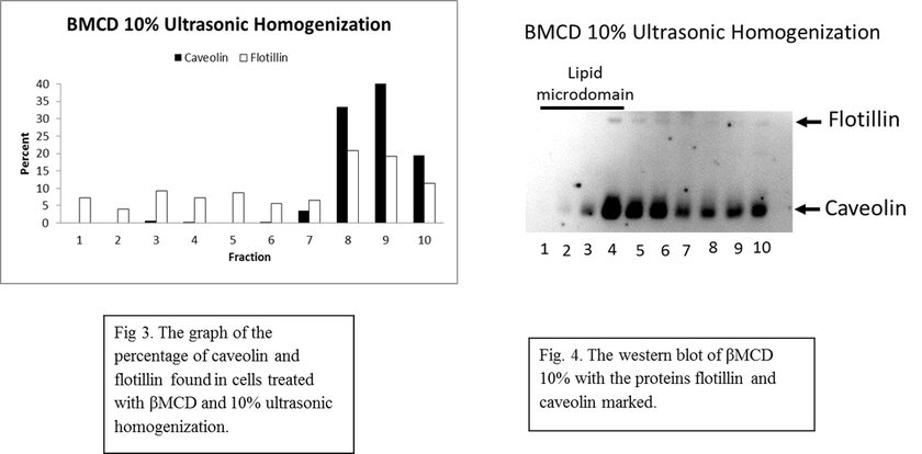
βMCD 10% ultrasonic homogenization had the highest percentage of caveolin and flotillin in fractions 7-10. There was no caveolin in fractions 1, 2, 4, 5, and 6 and minimal amounts in fraction 3. There was a lower percentage of flotillin in fractions 1-6. It was not until fractions 7-10 that there was a high percentage of the two proteins found within the cells (Fig. 3). The western blot of βMCD 10% demonstrated the same trend. Most of the proteins were found in the lower fractions (Fig. 4).
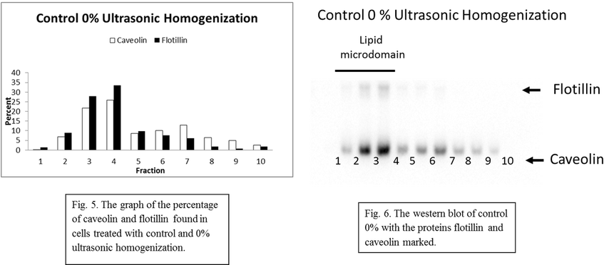
Control 0% ultrasonic homogenization had the highest percentage of caveolin and flotillin in fraction 3-7. Fractions 3 and 4 had the highest percentage proteins out of all ten fractions. The lower fractions had higher percentages of proteins when compared to the upper fractions (Fig. 5). The same results can also be seen when the western blot is examined (Fig. 6).
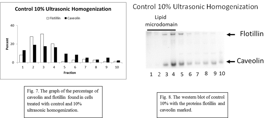
Control 10% ultrasonic homogenization had the highest percentage of caveolin and flotillin in fractions 1-4. There were a significantly lower percentage of these proteins in fractions 5-10. Fractions 2 and 3 had the highest percentage of proteins (Fig. 7). The western blot also demonstrated that there were a higher percentage of proteins in the lower fractions as compared to the upper fractions (Fig. 8).
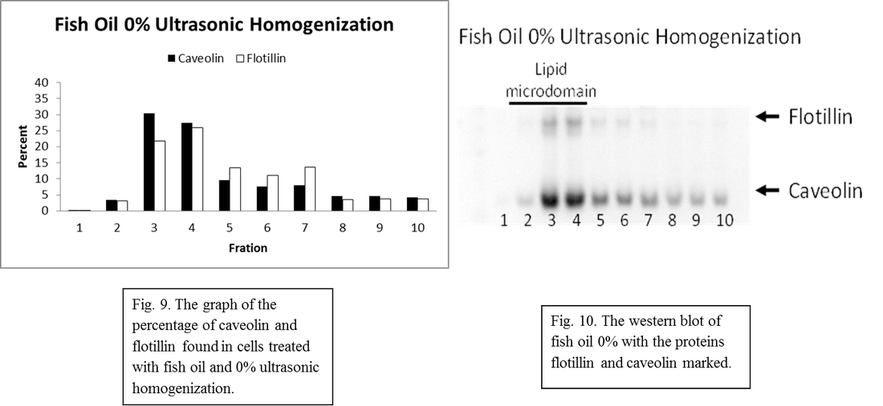
Fish oil 0% ultrasonic homogenization had the highest percentage of caveolin and flotillin in fractions 3 and 4. Fractions 5-7 also had relatively high percentages of the two proteins. Fractions 1, 2, 8, 9, and 10 all had low levels of caveolin and flotillin. The middle fractions had the highest percentage of the proteins (Fig. 9). This was also demonstrated with the western blot (Fig. 10).
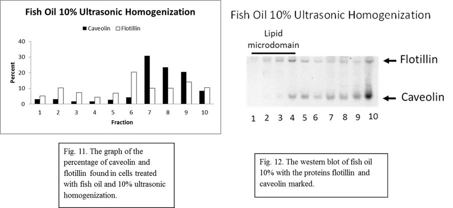
Fish oil 10% ultrasonic homogenization had significantly higher levels of caveolin and flotillin in the upper fractions. Fractions 6-10 had the highest percentage of the two proteins. The lower fractions such as 1-5 had low percentages of the two proteins. Both proteins had higher percentages in the upper fractions and less in the lower fractions (Fig. 11). The western blot also demonstrated a similar trend (Fig. 12).